We use cookies to make your experience better. To comply with the new e-Privacy directive, we need to ask for your consent to set the cookies.
MRI

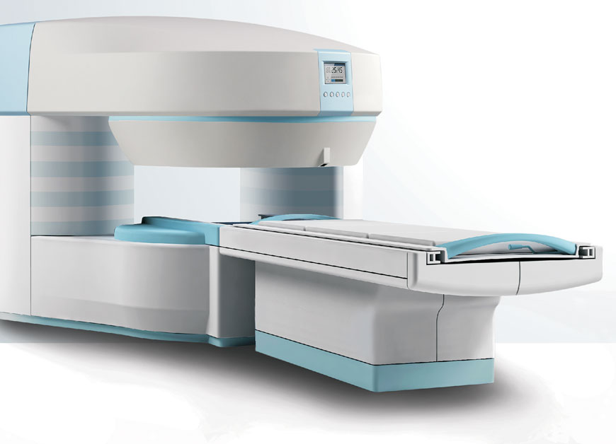
Magnetic resonance imaging (MRI) scanner also called nuclear magnetic resonance (NMR) imaging scanner. They use powerful magnets to polarise and excite hydrogen nuclei (single proton) in water molecules in human tissue, producing a detectable signal which is spatially encoded, resulting in images of the body.
MRI uses three electromagnetic fields: a very strong (on the order of units of teslas) static magnetic field to polarize the hydrogen nuclei, called the static field; a weaker time-varying (on the order of 1 kHz) field(s) for spatial encoding, called the gradient field(s); and a weak radio-frequency (RF) field for manipulation of the hydrogen nuclei to produce measurable signals, collected through an RF antenna. Like CT, MRI traditionally creates a two dimensional image of a thin "slice" of the body and is therefore considered a tomographic imaging technique. Modern MRI instruments are capable of producing images in the form of 3D blocks, which may be considered a generalisation of the single-slice, tomographic, concept.
Unlike CT, MRI does not involve the use of ionizing radiation and is therefore not associated with the same health hazards.

&width=200&height=200&quality=75)
&width=200&height=200&quality=75)
&width=200&height=200&quality=75)
&width=200&height=200&quality=75)
&width=200&height=200&quality=75)
&width=200&height=200&quality=75)
&width=200&height=200&quality=75)
&width=200&height=200&quality=75)
&width=200&height=200&quality=75)